- Have any questions?
- info@radiopaedia.ir

COVID19-Old Fibrosis and New Acute Pneumonitis
9th May 2020
COVID19_Patchy Consolidations
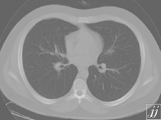
9th May 2020COVID19_Non-suggestive Sign for COVID-19 pneumonitis


Chest wall including ribs are normal without evidence of bone destruction. Small areas of ground glass appearance in left arterial para-hilar and little bit lower to hilum are non-suggestive or non-consistent sign for COVID-19 pneumonitis. With positive clinical findings please repeat CT scan of lung 3-5 days later. Aorta (ascending, arch, descending) and main brachiocephalic branches are normal. Pulmonary hilar structures, trachea, S.V.C and esophagus didn't show abnormality. Costo - phrenic sulci are clear. No pleural effusion is seen.

Radiopaedia is proudly powered by WordPress